Imaging of the dentofacial structures is an important diagnostic tool in comprehensive orthodontic treatment. In 1931, Broadbent pioneered the evaluation of dentofacial structures in three dimensions (3D) using two images taken in the sagittal (left to right) and frontal (back to front) views. Since then, with the advancement of radiological science, Sir Godfrey Hounsfield invented computed tomography (CT) in the early 1970s, starting the true era of 3D imaging. Modern medical-grade CT scans, with the assistance of computers, compile these 2D image slices and reformat them into true 3D images that are both stunning and vivid.
Wi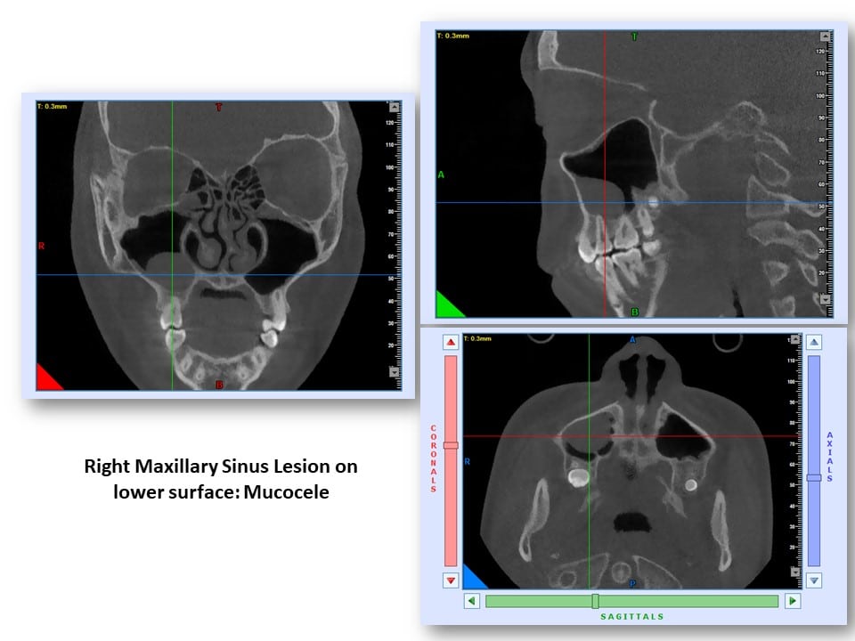
At OPDSF Orthodontics, we have the state-of-the art CBCT machine. The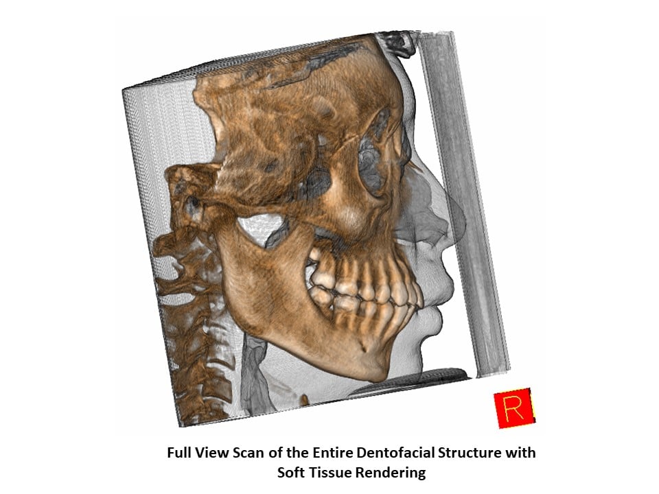
CBCT imaging allows us a more conservative and comprehensive view of the dentofacial structures, including the soft tissue structures that were previously difficult or impossible to evaluate. Some common applications of this new technology are listed below:
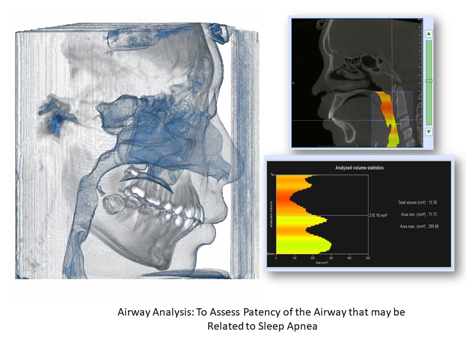
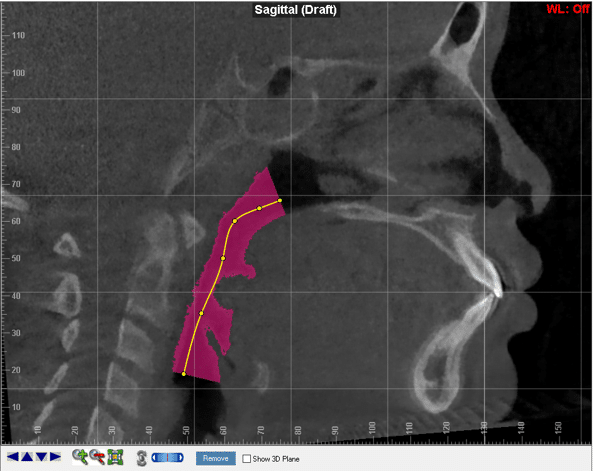
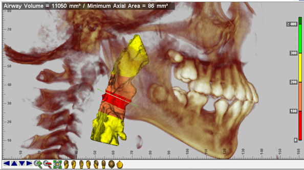
- Analyze the airway that may indicate the possibility of sleep apnea.
- Able to detect pattern and rate of facial growth, including any subtle asymmetry.
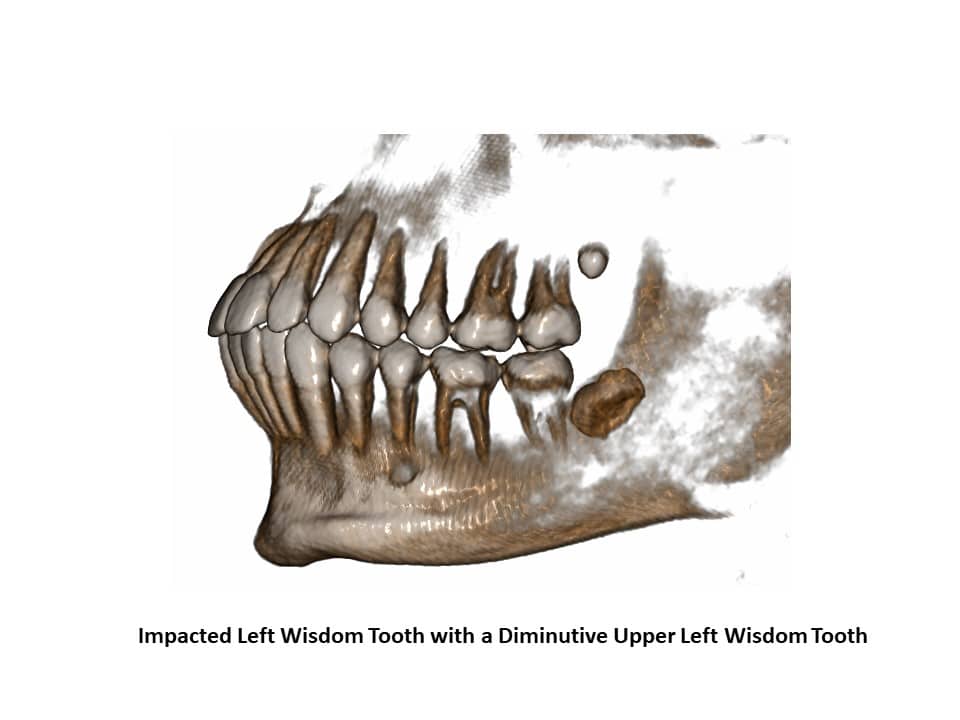
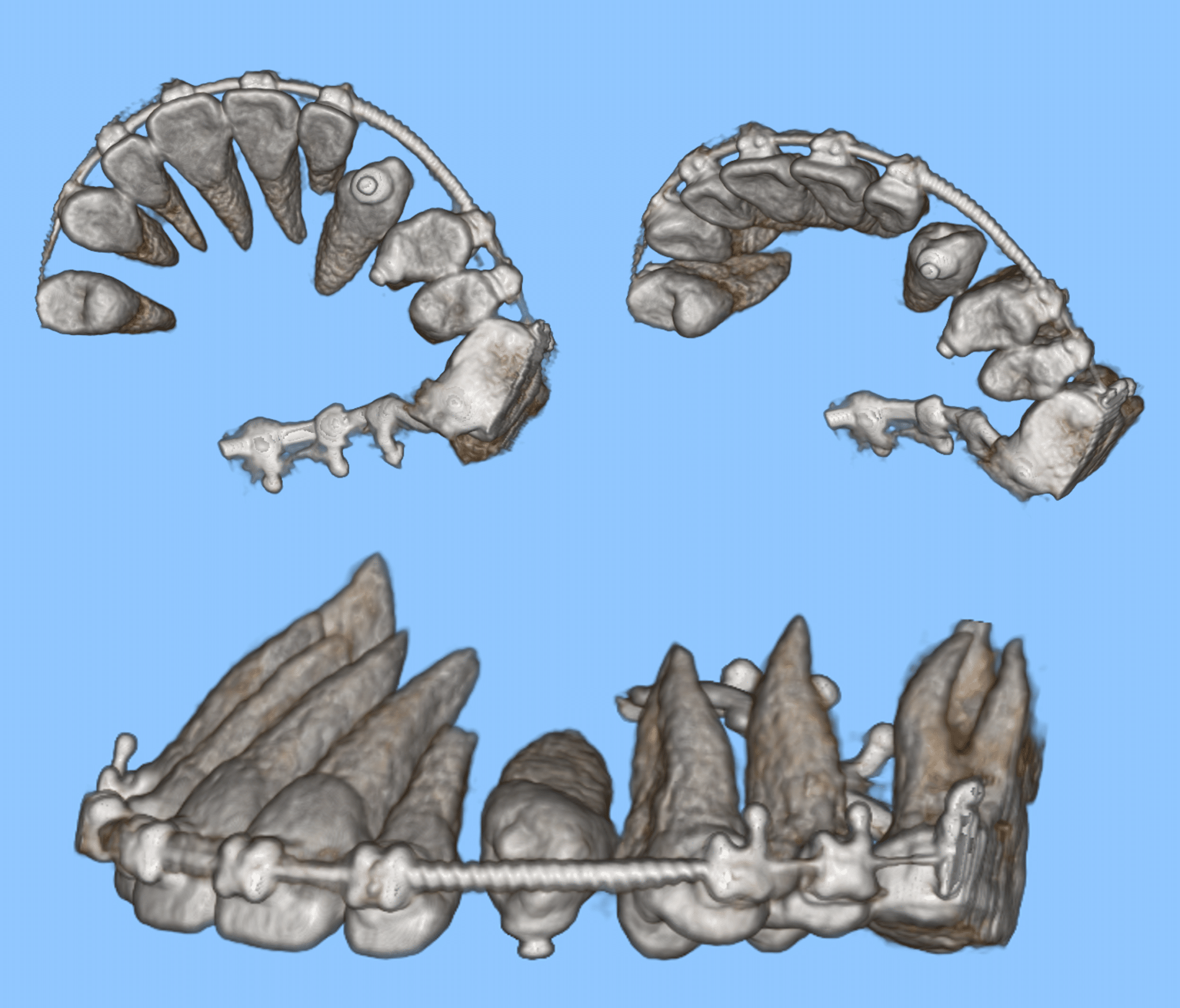
- Detect abnormalities, e.g. supernumerary teeth, impacted third molars (wisdom teeth), missing teeth, abnormal eruption path and primary failure of eruption.
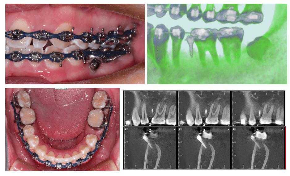
- Detect root foreshortening in other spatial planes that were not available conventionally.
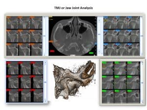
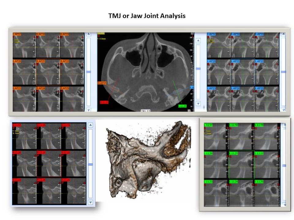
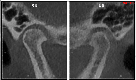
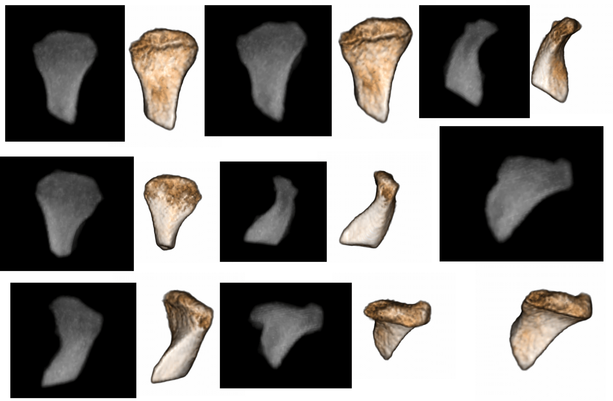
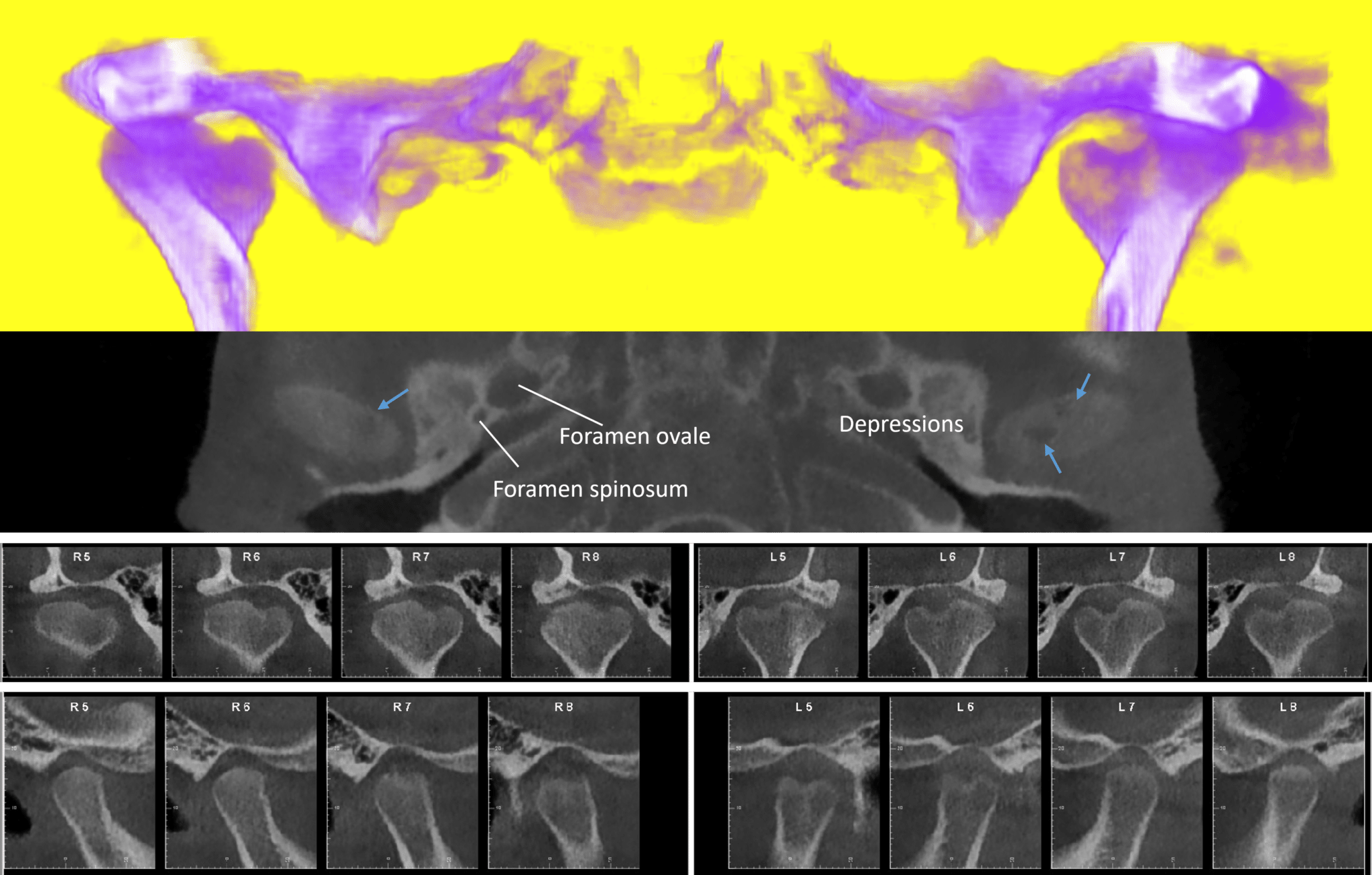
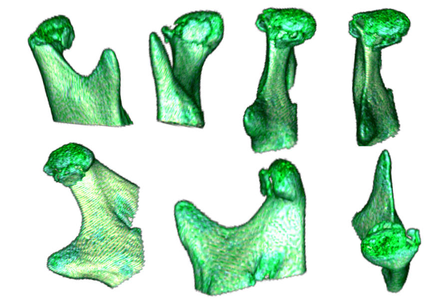
- Able to predict with confidence the limitations of orthodontic and surgical movements.Accurately evaluate the jaw joints (TMJs), e.g., jaw joint spaces, atrophy of condylar surfaces and osteoarthritis.
- Visualize unparalleled imagery of the entire dentofacial and soft tissue simultaneously.
- Determine the bone housing levels associated with gum problems (periodontitis).
- Asses damage to traumatized bone or teeth, e.g., root fracture.
- Evaluate the extent of periapical lesions associated with a root canal (endodontics).
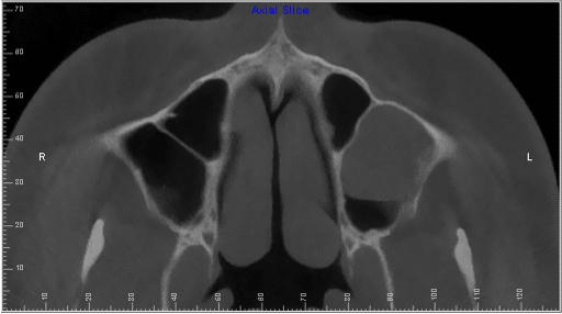
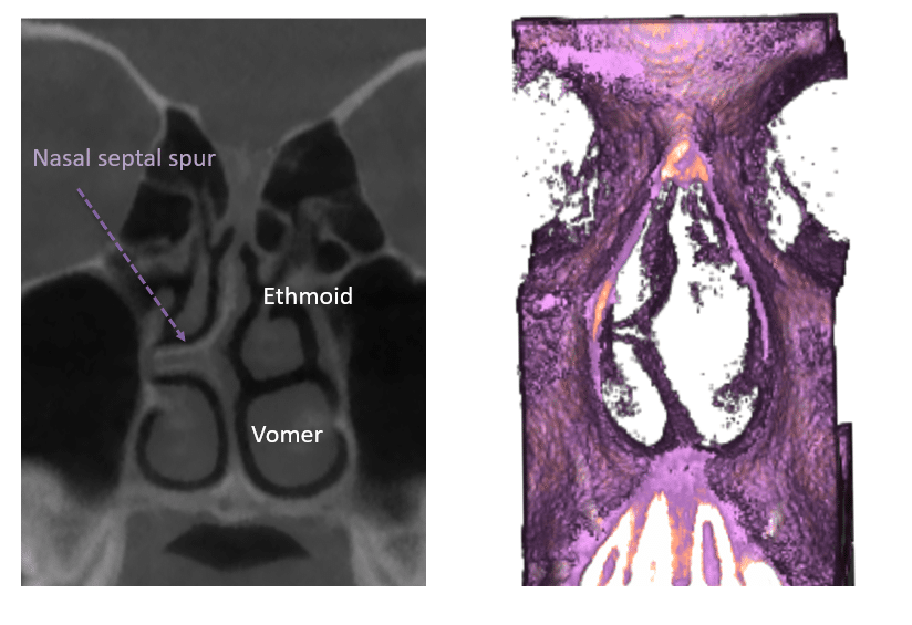
- Review the paranasal sinuses clearly, e.g., mucocele.
- Assess the quality and site of bone structure for future dental implants.
- Determine the maturation and growth potential of the dentofacial structures.
- Precisely determine the root positioning to allow accurate orthodontic correction.
- Accurately measure anatomical structures, e.g., thickness of upper palatal shelf, determination for skeletal mini pins and optimal sites for mini pin (Temporary Anchorage Device, TAD) placements.


